A glaucoma work-up is a comprehensive set of diagnostic tests used to detect glaucoma, a condition that causes damage to the optic nerve and can lead to permanent vision loss if left untreated. The goal of a glaucoma work-up is to assess the intraocular pressure (IOP), evaluate the optic nerve head, and detect any changes in the structure or function of the eye that may indicate glaucoma.
At Desai Eye Institute, the glaucoma work-up typically includes a combination of tests and examinations to thoroughly evaluate eye health. Key components of a glaucoma work-up include:
1. Intraocular Pressure Measurement (IOP) – This is usually done through tonometry, such as Applanation Tonometry, to check for elevated pressure inside the eye, which is a major risk factor for glaucoma.
2. Optic Nerve Head Examination – A detailed examination using a slit lamp or fundus camera to assess the optic disc for signs of damage, cupping, or other abnormalities.
3. Pachymetry – A measurement of corneal thickness, as thinner corneas can be associated with a higher risk of glaucoma.
4. Visual Field Testing – To detect any peripheral vision loss, a common early symptom of glaucoma.
5. Gonioscopy – A test used to examine the angle of the eye where the iris meets the cornea to assess whether it is open or closed, which helps determine the type of glaucoma.
6. OCT (Optical Coherence Tomography) – Imaging of the optic nerve and retinal nerve fiber layer to detect structural changes.
7. Fundus Fluorescein Angiography (if needed) – To assess the retinal vasculature and identify any abnormal blood flow or damage.
The results of these tests help determine the presence of glaucoma, monitor its progression, and guide treatment decisions to prevent further vision loss. Early detection and management are critical for preserving vision in glaucoma patients.
Medical Equipment Needed for Glaucoma Work-Up
Tonometry Device (Applanation Tonometer or Tonopen)
Slit Lamp (for detailed eye examination)
Pachymeter (for corneal thickness measurement)
Visual Field Analyzer (for detecting peripheral vision loss)
Gonioscope (for angle examination)
Optical Coherence Tomography (OCT) Machine
Fundus Camera (for optic nerve head and retinal imaging)
Fluorescein Dye (for angiography, if required)
Sterile Gloves and Disinfectants (for hygiene and equipment sterilization)
Patient Headrest and Chin Rest (for proper positioning during tests)
01
Make Appointment
If you need help booking an appointment, please call us.
02
Select Doctor
Choose a doctor from the list, review their profile, and select the most suitable one for your needs.
03
Get Consultation
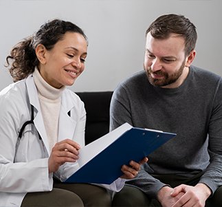
Book your onsite consultation and meet the doctor at the scheduled time.
04
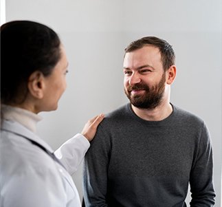
Get Cure & Relief
Experience complete care and relief as the doctor helps treat your condition effectively.