Professional
Clinic

4.9 (7.8k review)
Schedule Hours
Our Location

Desai Eye Institute & Research Center

Manharpark Society,
Subhanpura High Tension Line Road,
Near Vimalnath Cross Roads,
Santosh Nagar, Subhanpura,
Vadodara, Gujarat 390023
Appointments
Desai Eye Institute & Research Center offers regular OPD consultations and Emergency Medical Services (EMS), ensuring timely care for routine and urgent eye-related medical needs.

Book an Appointment:

Ph: 0265-2292266, 0265-3553505
M: +91 89804 92266

Who We Are
300+ Appointment <br> Booking Confirm for <br> this Week

300+ Appointment
Booking Confirm for
this Week

Who We Are

A Legacy of Excellence in Ophthalmic Care

Desai Eye Institute and Research Centre, located in Vadodara, Gujarat, stands at the forefront of advanced eye care, committed to transforming lives through unparalleled vision care. With decades of expertise, we have established ourselves as a trusted institution, offering comprehensive ophthalmic treatments that seamlessly blend cutting-edge medical technology with a compassionate, personalized approach.

Desai Eye Institute and Research Centre, located in Vadodara, Gujarat, stands at the forefront of advanced eye care, committed to transforming lives through unparalleled vision care. With decades of expertise, we have established ourselves as a trusted institution, offering comprehensive ophthalmic treatments that seamlessly blend cutting-edge medical technology with a compassionate, personalized approach.

At the core of our philosophy is a commitment to innovation, precision, and excellence. Equipped with state-of-the-art technology and driven by evidence-based practices, we consistently achieve exceptional outcomes. Our compassionate team of experts is dedicated to providing not just medical care, but a sense of trust and reassurance for every patient.

WHAT WE DO!

How it Helps You to Keep
Healthy

Applanation Tonometry
tab-img-1

Applanation Tonometry

Applanation tonometry is a diagnostic procedure to measure intraocular pressure (IOP), an essential test for glaucoma detection. It involves flattening a small area of the cornea with a gentle probe, ensuring accuracy in assessing the eye's pressure. Regular monitoring is critical for maintaining optimal eye health and preventing glaucoma-related damage to the optic nerve.

Foreign Body Removal
tab-img-1

Foreign Body Removal

Foreign body removal involves carefully extracting particles or debris from the eye that may cause irritation, injury, or infection. This procedure requires precision to prevent further damage to delicate ocular structures. After removal, patients are monitored for any signs of complications, such as infection or scarring, to ensure complete recovery and restore eye function.

Concretion Removal
tab-img-1

Concretion Removal

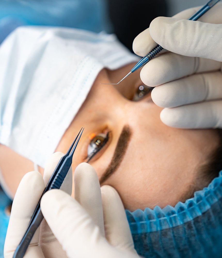
Concretion removal is a procedure to extract small, calcified deposits from the conjunctiva or eyelid. These deposits can cause irritation or discomfort. Removal is typically performed with a delicate surgical instrument under local anesthesia. The procedure is crucial for maintaining ocular health and preventing chronic irritation or infection from these deposits.

Dressing
tab-img-1

Dressing

Dressing after eye surgery or trauma serves to protect the eye and promote healing. A sterile dressing is applied to shield the eye from infection, reduce inflammation, and manage discomfort. Proper dressing care is essential to avoid complications like infection or improper healing, ensuring a faster recovery and optimal visual outcome.

Amniotic Membrane Grafting
tab-img-1

Amniotic Membrane Grafting

Amniotic membrane grafting is used to promote healing in cases of severe corneal injury or disease. The graft, derived from human placental tissue, is applied to the damaged area to facilitate tissue regeneration and reduce inflammation. This procedure enhances the repair of the cornea, improving the chances of vision restoration and minimizing scarring.

Corneal Tear Repair
tab-img-1

Corneal Tear Repair

Corneal tear repair is a delicate procedure aimed at restoring the integrity of the cornea after injury. Using sutures or other techniques, the tear is carefully closed to prevent infection and promote healing. Prompt treatment is critical to avoid complications, such as vision impairment, and to facilitate the healing of the corneal tissue.

Topography
tab-img-1

Topography

Corneal topography is a non-invasive imaging technique that maps the surface curvature of the cornea. It is essential for diagnosing and managing conditions like astigmatism, keratoconus, and pre-surgical planning for procedures like LASIK. Accurate topography ensures the proper treatment plan is followed for optimal visual outcomes and patient satisfaction.

Fundus Fluorescein Angiography
tab-img-1

Fundus Fluorescein Angiography

Fundus fluorescein angiography is a diagnostic tool used to examine the blood vessels in the retina and choroid. A fluorescent dye is injected into the bloodstream, allowing the physician to capture detailed images of the eye's vasculature. This procedure is vital for detecting retinal diseases like diabetic retinopathy, macular degeneration, and retinal vein occlusion.

Ophthalmic Ultrasonography
tab-img-1

Ophthalmic Ultrasonography

Ophthalmic ultrasonography uses sound waves to create images of the eye’s internal structures, particularly in cases where the view of the retina is obstructed. It is commonly used for diagnosing conditions like retinal detachment, tumors, or lens dislocation. This non-invasive technique provides valuable insight into ocular health and aids in treatment planning.

OCT (Optical Coherence Tomography)
tab-img-1

OCT (Optical Coherence Tomography)

Optical Coherence Tomography (OCT) is a non-invasive imaging technique that provides detailed cross-sectional images of the retina and optic nerve. OCT is essential for detecting early signs of retinal diseases, macular degeneration, and glaucoma. By providing high-resolution images, it aids in precise diagnosis and monitoring of disease progression, guiding treatment decisions.

Lasik Work
tab-img-1

Lasik Work

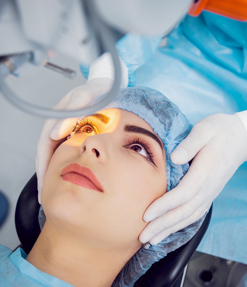
LASIK (Laser-Assisted in Situ Keratomileusis) is a refractive surgery used to correct vision issues like myopia, hyperopia, and astigmatism. It reshapes the cornea using a laser to improve light entering the eye. The procedure is quick, minimally painful, and offers fast recovery times, making it a popular choice for patients seeking to reduce their dependency on glasses or contacts.

Glaucoma Work-Up
tab-img-1

Glaucoma Work-Up

A glaucoma work-up involves a series of tests to assess the health of the optic nerve and measure intraocular pressure (IOP). It includes tonometry, visual field testing, and imaging techniques like OCT. Early detection of glaucoma is crucial for preventing vision loss, as the disease can progress without noticeable symptoms until significant damage has occurred.

Retina Laser
tab-img-1

Retina Laser

Retina laser treatment is used to treat a variety of retinal conditions, such as diabetic retinopathy and retinal tears. The laser helps to seal leaks in retinal blood vessels, repair tears, or prevent further damage to the retina. This minimally invasive procedure plays a vital role in preserving vision and preventing the progression of retinal diseases.

Intravitreal Injection
tab-img-1

Intravitreal Injection

Intravitreal injections involve the delivery of medication directly into the vitreous, the gel-like substance inside the eye. This treatment is commonly used for conditions like age-related macular degeneration (AMD), diabetic macular edema (DME), and retinal vein occlusion. The injections help reduce inflammation, control fluid leakage, and prevent further damage to the retina.

YAG Procedures
tab-img-1

YAG Procedures

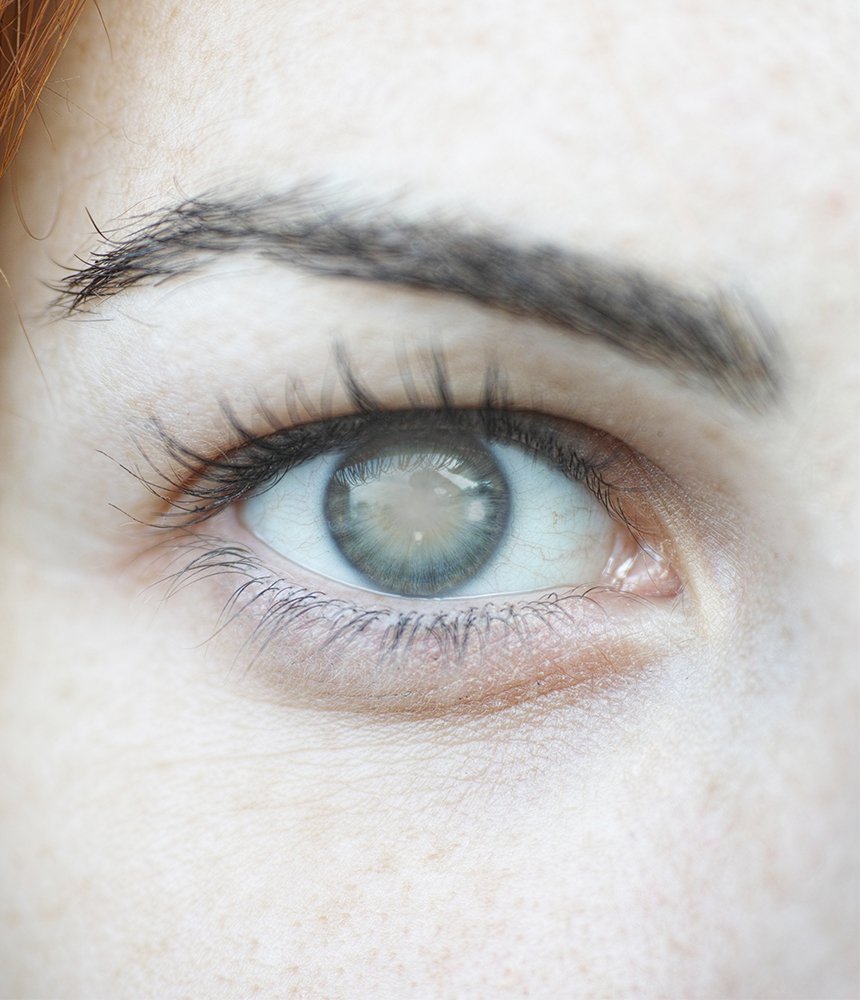
YAG (Yttrium-Aluminum-Garnet) laser procedures are commonly used to treat secondary cataracts, also known as posterior capsule opacification, after cataract surgery. The laser creates a small opening in the clouded capsule, restoring vision. YAG laser capsulotomy is a quick, painless procedure with minimal recovery time, improving the patient’s visual clarity.

Retinal Cryopexy
tab-img-1

Retinal Cryopexy

Retinal cryopexy is a procedure that uses freezing to treat retinal tears or detachment. The freezing causes scarring around the tear, helping to reattach the retina to the underlying tissue. It is often used in conjunction with other treatments, such as laser therapy, to prevent further retinal damage and preserve vision.

Posterior Sub-Tenon Injection
tab-img-1

Posterior Sub-Tenon Injection

Posterior sub-tenon injections deliver medication directly into the space beneath the Tenon’s capsule, surrounding the eye. This method is used to treat conditions such as uveitis, diabetic retinopathy, and macular edema. It allows for targeted delivery of steroids or other medications to reduce inflammation and prevent vision-threatening complications.

Surgeries
tab-img-1

Surgeries

Eye surgeries encompass a range of procedures aimed at correcting or treating ocular diseases, trauma, and conditions. Surgeries can include cataract removal, glaucoma procedures, or retinal surgery. Each procedure is performed with precision to improve or restore vision, with careful post-operative care to ensure optimal recovery and minimize complications.

Pterygium
tab-img-1

Pterygium

Pterygium is the growth of tissue on the conjunctiva, often extending onto the cornea. Treatment involves surgical removal of the growth, particularly when it affects vision or causes discomfort. Post-operative care is crucial to ensure proper healing and prevent recurrence. Surgery is effective in restoring comfort and preventing further vision obstruction.

Supra-Tarsal Injection
tab-img-1

Supra-Tarsal Injection

Supra-tarsal injections deliver medication into the upper eyelid, beneath the tarsal plate, to treat inflammatory or allergic conditions like conjunctivitis or uveitis. This method ensures targeted delivery of drugs such as corticosteroids, providing effective relief and reducing inflammation without affecting other parts of the eye.

Retinal Surgeries
tab-img-1

Retinal Surgeries

Retinal surgeries are performed to treat conditions affecting the retina, including retinal detachment, diabetic retinopathy, and macular degeneration. These procedures may involve laser therapy, vitrectomy, or retinal reattachment techniques to preserve or restore vision. Prompt intervention is critical to prevent permanent damage and maintain ocular health.

Corneoscleral Tear Repair
tab-img-1

Corneoscleral Tear Repair

Corneoscleral tear repair is performed to treat severe eye injuries involving both the cornea and sclera. The procedure involves suturing the torn tissue back together to restore the integrity of the eye and prevent complications such as infection, prolapse, or vision loss. Early surgical intervention is essential for a positive outcome.

Lid Repair
tab-img-1

Lid Repair

Lid repair is a surgical procedure performed to treat eyelid injuries, such as lacerations or tears. The goal is to restore both the function and appearance of the eyelid, ensuring proper eyelid closure and preventing complications like corneal exposure or infection. Timely treatment helps avoid long-term damage or scarring.

Enucleation/Evisceration
tab-img-1

Enucleation/Evisceration

Enucleation and evisceration are surgical procedures performed to remove an eye that is severely damaged or diseased. Enucleation involves the complete removal of the eye, while evisceration involves removing the inner contents of the eye, leaving the outer shell intact. Both procedures are performed to relieve pain and prevent further complications.

Cyclocryopexy
tab-img-1

Cyclocryopexy

Cyclocryopexy is a procedure that uses freezing to treat certain types of glaucoma or retinal conditions. The freeze is applied to the ciliary body, which produces intraocular fluid, reducing the pressure within the eye. This technique is often used when other treatments have failed, helping to control intraocular pressure and prevent further vision loss.

Meibography
tab-img-1

Meibography

Meibography is an imaging technique used to evaluate the meibomian glands in the eyelids, which are responsible for producing the oily layer of the tear film. It helps diagnose dry eye disease, meibomian gland dysfunction, and other related conditions. Early detection allows for timely intervention to prevent long-term discomfort and visual impairment.

Contact Us

Make an appointment.

    4.82

    rating-black
    2,488 Rating
    Welcome to Desai Eye Institute & Research Centre!

    We’re open 24/7.

    For OPD appointments:

    Ph: 0265-2292266, 0265-3553505

    M: +91 89804 92266

    0265-229-2266

    info@desaieyeinstitute.com

    Homepage 07